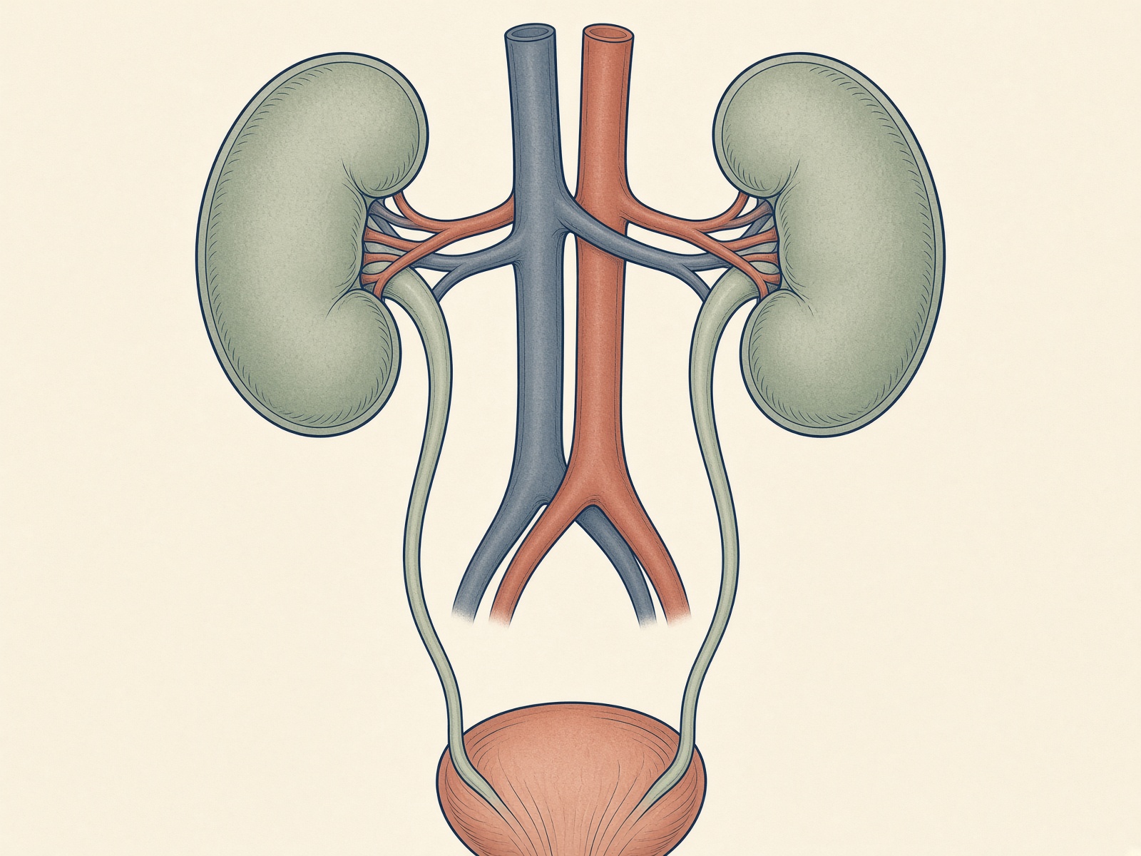
A kidney stone is a hard deposit that forms when urine is too concentrated with minerals — calcium, oxalate, uric acid, phosphate, or cystine. The stone itself can sit quietly in the kidney for months. The famous pain starts when a fragment drops into the ureter and the tube spasms around it.
Pain usually starts when a fragment leaves the kidney and enters the ureter.
Symptoms worth taking seriously
Sharp, wave-like pain in the flank, side, lower abdomen, or groin (renal colic)
Pain that migrates as the stone moves
Pink, red, or brown urine (blood)
Urgent, frequent, or burning urination if the stone is near the bladder
Nausea and vomiting from the shared nerve pathways with the gut
Fever or chills — that is infection until proven otherwise
Source: NIDDK, Symptoms & Causes of Kidney Stones.
The five crystal types
Calcium oxalate — the majority
Most first stones in adults. They form when urine is high in calcium and oxalate, low in volume, or low in citrate. Paradox: cutting dietary calcium often raises oxalate absorption and makes these worse. Eat calcium with meals instead.
Calcium phosphate
More common when urine is alkaline. Prevention still leans on fluids and sodium control. Citrate therapy can raise pH, so these stones need a clinician watching the chemistry — alkali is not automatic.
Uric acid
Form in acidic urine, often with gout, metabolic syndrome, or a heavy animal-protein pattern. They can sometimes be dissolved with prescription alkali that raises urine pH. Imaging may miss them on a plain X-ray.
Struvite (infection stones)
Grow after certain urinary infections. They can become large staghorn stones quickly. Antibiotics and removal are the treatment; lemon water is not.
Cystine
A genetic transport disorder. These patients drink heroic volumes and need specialty meds. If you have cystinuria, this general site is a starting point only.
Why they form
Urine is a crowded solution. When water is low, or promoters (calcium, oxalate, uric acid, sodium-driven calcium leak) outrun inhibitors (citrate, magnesium, volume), crystals nucleate on a tiny nidus — often a calcium-phosphate plaque on a papilla, called a Randall’s plaque.
Modifiable risks the research keeps repeating: low fluid intake, high sodium, high animal protein, a low-calcium diet, high oxalate load, obesity, hot work or desert climate, and holding urine for hours. Family history and certain meds (topiramate, some diuretics, excess vitamin C in some people) stack on top.
How doctors confirm it
Non-contrast CT is the gold-standard snapshot. Ultrasound avoids radiation and is useful in pregnancy. A stone analysis after you catch one is the single most useful lab you can bring to a prevention visit. A 24-hour urine collection then shows whether the problem is volume, calcium, oxalate, citrate, uric acid, sodium, or pH.